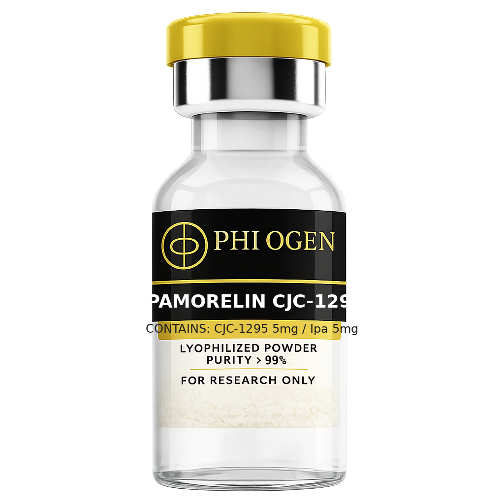
CJC-1295: GHRH Analog with Drug Affinity Complex Technology
CJC-1295 is a synthetic analog of growth hormone-releasing hormone (GHRH), the hypothalamic peptide responsible for stimulating somatotroph cells in the anterior pituitary to secrete growth hormone. Native GHRH(1-44) and its truncated biologically active form GHRH(1-29) both carry a serine at position 2 that is rapidly cleaved by the serum enzyme dipeptidyl peptidase IV (DPP-IV), giving endogenous GHRH a circulating half-life of only 6–7 minutes. The first-generation research analog sermorelin (GHRH 1-29 amide) extends this modestly but still degrades within approximately 30 minutes following subcutaneous administration.
CJC-1295 addresses this limitation through Drug Affinity Complex (DAC) technology — a proprietary modification in which a maleimidopropionic acid (MPA) linker is conjugated to the C-terminus of GHRH(1-29). The maleimide group reacts via Michael addition with the free thiol of cysteine-34 on circulating human serum albumin, forming a covalent thioether bond in vivo. Because albumin has a half-life of approximately 19–20 days and is largely excluded from renal filtration due to its 66.5 kDa molecular weight, the CJC-1295/albumin adduct inherits a dramatically extended pharmacokinetic profile. Published research in healthy adults demonstrated that a single subcutaneous injection of CJC-1295 produced measurable growth hormone-releasing activity persisting for 6–8 days, in stark contrast to the minutes-scale activity of unmodified GHRH.
At the receptor level, CJC-1295 retains full agonist activity at the GHRH receptor (GHRHR), a Gs-protein-coupled receptor expressed on pituitary somatotroph cells. GHRHR activation elevates intracellular cyclic AMP (cAMP), activates protein kinase A (PKA), and ultimately triggers vesicular exocytosis of pre-formed GH granules while simultaneously upregulating GH gene transcription. The albumin-bound DAC form engages the receptor with slightly reduced potency per molar concentration compared to free GHRH(1-29), but this is more than compensated by the sustained plasma availability. Importantly, somatostatin-mediated feedback is preserved — the hypothalamic-pituitary axis continues to modulate pulsatile release, so CJC-1295 amplifies rather than replaces endogenous GH regulation.
Ipamorelin: Fifth-Generation Selective Growth Hormone Secretagogue
Ipamorelin is a pentapeptide (Aib-His-D-2-Nal-D-Phe-Lys-NH2) classified as a growth hormone secretagogue (GHS) and recognized as a selective ghrelin receptor (GHSR1a) agonist. The GHSR1a is a Gq-protein-coupled receptor that, when activated, mobilizes intracellular calcium stores and stimulates PKC-mediated signaling in somatotroph cells. Unlike ghrelin itself — the endogenous GHS-R1a ligand — ipamorelin was engineered to maximize GH selectivity while minimizing off-target hormonal effects.
The development arc of GHRPs is instructive here. First-generation secretagogues (GHRP-6, hexarelin) produced dose-dependent increases in cortisol and prolactin alongside GH, as the GHSR1a is expressed on adrenal and lactotroph cells in addition to somatotrophs. GHRP-2 reduced but did not eliminate these effects. Ipamorelin, characterized in the late 1990s by Novo Nordisk researchers, demonstrated a selectivity profile in which equimolar doses that produced robust GH pulses generated no statistically significant elevations in ACTH, cortisol, or prolactin in preclinical rodent studies — distinguishing it as a fifth-generation GHS.
Mechanistically, ipamorelin operates through two converging pathways. It directly stimulates GHSR1a on pituitary somatotrophs and also acts at the hypothalamic level to suppress somatostatin release, thereby removing the inhibitory brake on GH secretion. This dual action is partly shared with ghrelin but ipamorelin's selectivity for the pituitary GHSR1a over hypothalamic somatostatin neurons appears to be responsible for its cleaner hormonal profile. The half-life of ipamorelin following subcutaneous administration is approximately 2 hours, making it well-suited to periodic dosing protocols designed to mimic physiological GH pulsatility when used in conjunction with a longer-acting GHRH analog.
Synergistic Rationale: Amplitude and Frequency of GH Pulses
The combination of CJC-1295 and ipamorelin is premised on complementary and synergistic mechanisms at the pituitary level. GHRH analogs primarily increase the amplitude of individual GH pulses — they potentiate the magnitude of GH release per somatotroph cell activation event. GHRPs, by contrast, increase the frequency of GH pulse initiation, partly by suppressing hypothalamic somatostatin secretion and partly by independent somatotroph stimulation via GHSR1a. When both receptor systems are activated simultaneously, research in rodent and primate models consistently shows supraadditive GH release relative to either agent alone.
A key study by Bowers and colleagues demonstrated that co-administration of GHRH and GHRP-6 produced GH pulses of 2–5 fold greater amplitude than either peptide administered individually at equivalent molar doses. Ipamorelin, substituted for GHRP-6, recapitulates this synergy without the cortisol co-secretion issue. The mechanistic basis involves convergent intracellular signaling: GHRHR activation elevates cAMP/PKA while GHSR1a activation elevates DAG/PKC. Both converge on phospholipase C activation and IP3-mediated calcium release from the endoplasmic reticulum, and the combined intracellular calcium signal exceeds the threshold achievable by either pathway in isolation.
From a translational research perspective, the combination is designed to restore a youthful GH secretion pattern. Age-related GH decline (somatopause) is characterized by decreased pulse amplitude and frequency, reduced GHRH sensitivity, and elevated somatostatin tone. CJC-1295 addresses the amplitude deficit by providing sustained GHRH receptor stimulation over days, while ipamorelin injections timed to coincide with natural GH peaks (typically at sleep onset and post-exercise) augment pulse frequency. The net result in controlled rodent studies is normalization of 24-hour integrated GH AUC toward values observed in younger cohorts.
GH Pulse Physiology: Somatotrophs, Somatostatin, and Pulsatility
Understanding the physiological context for CJC-1295/ipamorelin research requires appreciation of the complex neuroendocrine architecture governing GH secretion. Somatotroph cells account for approximately 35–40% of anterior pituitary cells and store GH in dense secretory granules. GH secretion is pulsatile in all mammals studied, with humans exhibiting 8–14 discrete GH pulses per 24-hour period, most clustered in the early hours of nocturnal sleep. Between pulses, circulating GH is often undetectable, distinguishing pulsatile from the tonic (continuous low-level) secretion pattern seen with certain pituitary pathologies.
The pulse generator is a dual-oscillator system involving two hypothalamic peptides with opposing effects: GHRH promotes GH release, and somatostatin (somatotropin release-inhibiting factor, SRIF) suppresses it. GHRH neurons in the arcuate nucleus and somatostatin neurons in the periventricular nucleus fire in alternating bursts, creating the pulsatile output. The GHRH:SRIF ratio at the pituitary determines pulse timing and amplitude. Ghrelin, produced primarily in gastric oxyntic cells but also in hypothalamic neurons, provides a third regulatory input that amplifies GHRH signaling and suppresses SRIF.
Critically, pulsatile GH secretion is not merely a physiological curiosity — it is required for appropriate downstream signaling. Hepatic GH receptor signaling is desensitized by continuous GH exposure through receptor internalization and upregulation of suppressors of cytokine signaling (SOCS proteins). Pulsatile GH maintains receptor sensitivity by allowing receptor recycling during trough periods. This explains why continuous GH infusion in research models produces a markedly blunted IGF-1 response compared to pulsatile delivery at the same total dose, and why research protocols using GHS combinations are designed to respect the natural pulse architecture rather than override it.
The IGF-1 Axis: Hepatic Production and Anabolic Signaling
The primary systemic mediator of GH's anabolic effects is insulin-like growth factor 1 (IGF-1), a 70-amino acid single-chain polypeptide produced predominantly in the liver following GH receptor activation. Hepatocyte GH receptor (GHR) engagement activates the JAK2/STAT5b signaling cascade, which translocates to the nucleus and drives transcription of the IGF1 gene. IGF-1 is released into the circulation where approximately 75% is bound in a ternary complex with IGF binding protein 3 (IGFBP-3) and an acid-labile subunit (ALS), creating a large 150 kDa complex that extends IGF-1 half-life from approximately 12 minutes (free) to 12–15 hours (ternary complex).
IGF-1 acts on the type 1 IGF receptor (IGF-1R), a transmembrane receptor tyrosine kinase expressed in virtually all tissues. Upon ligand binding, IGF-1R undergoes autophosphorylation and recruits insulin receptor substrate proteins (IRS-1, IRS-2), initiating two major downstream cascades: the PI3K/AKT/mTOR pathway (mediating protein synthesis, glucose uptake, and cell survival) and the RAS/MAPK/ERK pathway (mediating cell proliferation and differentiation). In skeletal muscle, IGF-1/IGF-1R signaling through AKT-mTORC1 activates ribosomal S6 kinase (S6K1) and inhibits the eIF4E-binding protein 4E-BP1, collectively promoting mRNA translation and protein accretion.
GH also has direct anabolic effects independent of IGF-1, particularly on lipolysis (via adipose GHR activation of hormone-sensitive lipase) and on glucose metabolism. However, GH-deficient individuals have substantially reduced circulating IGF-1, and restoration of GH pulsatility through GHRH/GHS combinations consistently elevates serum IGF-1 in research models, serving as a convenient pharmacodynamic biomarker for GH-axis activation. The mean IGF-1 elevation observed in published CJC-1295 monotherapy studies was 2–3 fold above baseline, sustained over the multi-day dosing window.
Body Composition Research in GH-Deficient and Aged Rodent Models
GH-deficient rodent models — most notably the dw/dw dwarf rat and the GH-deficient hypophysectomized rat — have been instrumental in characterizing the body composition effects of GHRH analogs and GHS combinations. These models exhibit the hallmark features of adult GH deficiency: increased visceral adipose tissue mass, reduced lean body mass, decreased bone mineral density, and impaired exercise tolerance. Restoration of GH pulsatility via pharmacological means in these models reverses many of these phenotypic features, providing mechanistic evidence for GH-axis interventions.
In studies using GHRH agonists, GH-deficient rodents showed significant increases in lean body mass (quantified by dual-energy X-ray absorptiometry, DEXA) and reductions in epididymal and retroperitoneal fat pad weights after 4–8 weeks of treatment. The mechanism involves both direct GH-mediated lipolysis and IGF-1-mediated muscle protein synthesis. GH activates hormone-sensitive lipase in adipocytes and promotes fatty acid oxidation via peroxisome proliferator-activated receptor alpha (PPARα) upregulation in liver and muscle. The net shift in fuel utilization from carbohydrate to fatty acid oxidation — reflected in reduced respiratory quotient (RQ) in metabolic cage studies — accounts for the fat mass reduction independent of caloric intake changes.
Aged rodent models (24-month-old rats) partially phenocopy GH deficiency despite intact pituitary anatomy, reflecting age-related somatostatin hypersecretion and reduced somatotroph sensitivity to GHRH. CJC-1295/ipamorelin combination treatment in aged rodents in preclinical studies has been reported to produce GH pulse restoration with downstream improvements in grip strength, muscle cross-sectional area, and adipose tissue distribution compared to vehicle-treated age-matched controls. These findings underscore the research hypothesis that pharmacological restoration of GH axis activity may attenuate age-related body composition changes.
Sleep Quality Research: GH Pulsatility and Slow-Wave Sleep
The relationship between GH secretion and sleep architecture is bidirectional and well-established. The largest GH pulse of the 24-hour cycle in humans occurs within the first hour of sleep onset and is temporally coincident with the first episode of slow-wave sleep (SWS, stages N3/N4). SWS is characterized by electroencephalographic delta waves (0.5–4 Hz), cortical synchronization, and reduced hypothalamic somatostatin tone. The permissive reduction in somatostatin during SWS, combined with the nocturnal peak of hypothalamic GHRH, creates optimal conditions for maximal pituitary GH output.
GHRPs including ipamorelin administered at sleep onset have been investigated for their effects on sleep architecture in preclinical models. GHRP-6 and GHRP-2 infusions in rats were found to increase the proportion of SWS and reduce REM sleep density, consistent with the hypothesis that endogenous ghrelin acts as a sleep-promoting signal. Ghrelin receptor knockout mice show fragmented sleep and reduced SWS, implicating the GHSR1a in normal sleep architecture maintenance. Ipamorelin's selectivity for GHSR1a without significant ghrelin-like peripheral effects makes it a useful research tool for isolating central sleep-regulatory effects of GHS receptor activation.
The research implication for CJC-1295/ipamorelin combination work is that administration timed to sleep onset may recruit natural GH-SWS coupling mechanisms to produce both a pharmacological and physiological GH pulse simultaneously. Whether the combined peptide-driven and sleep-driven pulse produces additive or merely overlapping secretory events depends on the degree to which somatostatin suppression by ipamorelin persists throughout the SWS episode. Investigations using continuous blood sampling in rodent telemetry models have begun to characterize this temporal interaction.
Recovery, Collagen Synthesis, and Connective Tissue Research
Beyond muscle and fat, GH axis research has extensively characterized the role of GH and IGF-1 in connective tissue biology. Type I and type III collagen synthesis in fibroblasts is GH-dependent, with GH deficiency associated with reduced collagen turnover, thinner skin, and impaired tendon mechanical properties in animal models. IGF-1 directly stimulates fibroblast proliferation and collagen gene (COL1A1, COL1A2, COL3A1) transcription via IGF-1R/PI3K/Akt signaling, and GH augments this through additional STAT5b-mediated transcriptional regulation.
In tendon research, mechanical strength is critically dependent on collagen fibril density and cross-linking. Aging and GH deficiency are associated with reduced fibril diameter and collagen cross-link maturity. Rodent studies with GH replacement showed increased tendon wet weight, collagen content (hydroxyproline assay), and failure load in Achilles tendon specimens after 8 weeks of treatment. GHRH analog studies have not yet characterized tendon endpoints as completely, but given that CJC-1295 drives IGF-1 elevation comparable to exogenous GH administration in pharmacokinetic studies, similar connective tissue effects are mechanistically anticipated.
Wound healing models in GH-deficient rodents demonstrate delayed epithelialization and reduced tensile strength at the wound site compared to GH-replete controls, with GHRH treatment partially correcting both parameters. The mechanism involves GH-driven fibroblast migration and IGF-1-driven fibroblast proliferation and extracellular matrix deposition. Growth plate cartilage research in juvenile hypophysectomized animals also documents GH-dependent chondrocyte proliferation and IGF-1-dependent matrix synthesis, providing a framework for understanding the multiple connective tissue outcomes relevant to CJC-1295/ipamorelin combination investigation.